


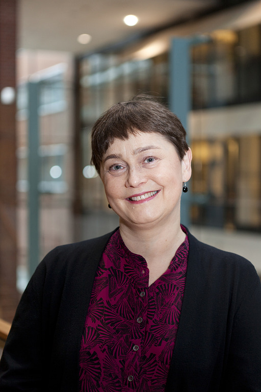
New Aalto Distinguished Professor Riitta Salmelin traces the brain’s own ‘fingerprint’
Riitta Salmelin believes that her field of brain imaging has matured to a stage where questions ignored in the early days can now be addressed

Over the past few decades, real-time tracking of cortical current flow and accurate localisation of blood oxygenation changes have offered complementary windows to the functional architecture of language in the human brain (MEG, fMRI; local activation, connectivity patterns).
The neuroimaging domain has reached its first level of maturity: we now know how to reliably measure and quantify different types of neuroimaging signals, we know that those signals show good intraindividual reproducibility but notable inter-individual variability in language tasks and, phenomenologically, we know what type of group-level functional effects to expect in a large variety of experimental conditions. To reach the next phase in the neuroscience of language, it is essential
To reach these goals, we combine careful experimental design and data collection and analysis with computationally explicit models and machine learning approaches.

Riitta Salmelin believes that her field of brain imaging has matured to a stage where questions ignored in the early days can now be addressed

Studies show disorder is linked to the left cerebral hemisphere and that confidence can help kids overcome difficulties

AI-Mind is a 5-year project funded by Horizon 2020, with the goal of facilitating a paradigm shift in clinical practice of mild cognitive impairment. A team of Aalto University and HUS Helsinki University Hospital researchers are involved in the project

Researchers are planning to investigate severe respiratory problems in coronavirus patients with EEG and machine learning, to help to predict the need of intensive care treatment of other COVID19 patients.

The way that speech processing differs from the processing of other sounds has long been a major open question in human neuroscience.

Partnered with machine learning, brain scans reveal how people understand objects in our world.

The awards is a well know prestige among international brain imaging researchers