






Health x technology: Easier, safer knee surgery with 3D cameras

Osteoarthritis is one of the most common disorders in ageing populations. Around 3.5 million hip or knee replacement operations are done every year in the OECD countries alone – more than 5,000 knee replacements each day.
A knee replacement operation typically takes just over an hour. You could almost say that new knees are flying off the assembly line! But industrial assembly lines use special equipment to run safely and efficiently. A 3D camera locates a car part so that a robot can pick it up and install it correctly. 3D cameras also scan logs as they move towards a saw. Machine vision helps industry automate its processes.
But joint replacement surgery doesn’t use 3D cameras. Ville Pitkänen first wondered why nearly 20 years ago. At the time, he was working on his master’s thesis, which included elements of computer-assisted orthopaedic surgery. After that, he went to work for a company supplying robots for joint replacement operations. Now, he’s part of the Aalto University ORCA project, which started last year.
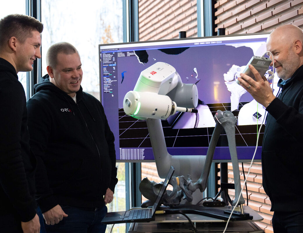
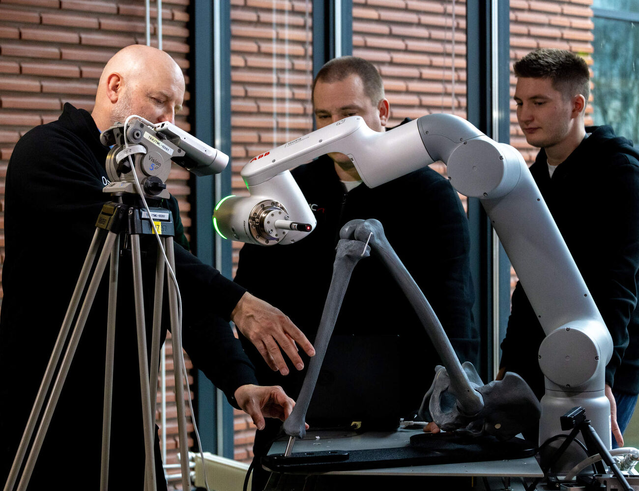
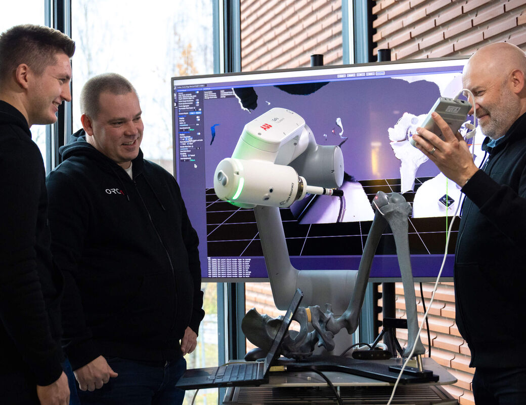
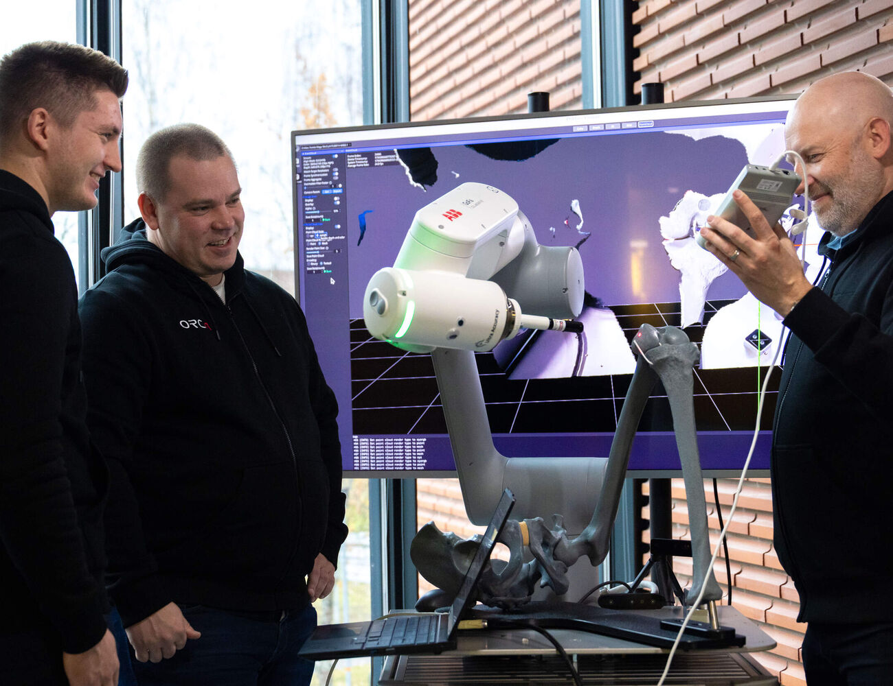
‘We’re developing a navigation system for joint replacement surgery in which a 3D camera sees the bone and tracks its movement. We’ve started developing it for knee replacement operations,’ Pitkänen says.
Pitkänen acknowledges that knee replacements already produce good results with current methods. Roughly speaking, almost 90 percent of patients are satisfied with the outcome. In cases where pain or stiffness in the knee persists, it’s not necessarily because the operation wasn’t precise enough. But improving precision isn’t the research group's main objective. Simplicity is.

‘We want to eliminate all the extra steps and equipment that take time, increase risks, and ultimately add costs. Our vision is that the navigation system will start tracking the bone automatically and help the surgeon as soon as the operation begins.’
Replacing cumbersome tracking markers
To Pitkänen, the main problem with current operations is the reflective markers attached to the bones to help locate them. After having followed hundreds of operations, he has developed a rather colourful vocabulary when it comes to describing his frustration with these markers. To understand why, we need to know what happens in the operating theatre.
In a conventional knee replacement operation, the surgeon plans the procedure based on x-ray images. After opening the knee, they determine where and at what angle the thigh bone and shin bone should be cut, and then they position mechanical cutting guides using anatomical landmarks.
The surgeon works on the bone like a fine carpenter, and this approach has produced good results since the 1990s. But each year, more surgeons are using a robotic arm to help position the joint replacement more precisely.
The robot can use a three-dimensional model based on computed tomography (CT) scans, but it can also operate without one. What’s essential is that the robot knows where the bones are at the beginning of the operation and how they move during the procedure. That’s where the reflective markers come in.
The markers are palm-sized, cage-like structures with spheres that reflect infrared light from a camera. The surgeon screws two or three markers into the bones and calibrates the navigation system by touching the bone at several dozen points with a sharp-tipped tool that also has a marker.
‘Calibration, which ensures the accuracy of the measurements, takes several minutes. And if the markers shift, the entire calibration process has to be done again,’ Pitkänen says. He explains that using the markers can easily make an operation take about 15 minutes longer even though the work on the bone goes faster with the robot’s help.
‘There’s a link between the length of an operation and the risk of infection. The risk of infection also increases when extra instruments are used and extra holes are drilled into the bone. Some of the markers are also attached through the skin,’ he explains.
Pitkänen wants to remove this fiddly process from the surgeon’s work and replace it with a 3D camera that wouldn’t need calibration.
Machine vision recognises the bone
Pitkänen says that robots are used in around 10–20 percent of knee replacement operations worldwide. They were first used in Finland in 2021. Australia has been a pioneer in the field, with robots used in 42 percent of knee operations in 2024.
Pitkänen believes that these figures will continue to grow. The precision offered by a robot isn’t necessarily needed for a good outcome, but it can help avoid unsuccessful results in exceptional cases. ‘Quite a lot of people use cruise control and lane assist in their cars, even though they could get to their destination without them,’ he says.
With the increasing use of robots, there’s a growing need for an easy-to-use and accurate navigation system, says Pitkänen. Greater precision isn’t the main goal, but precision will improve when reflective markers are replaced with a 3D camera. ‘If you have four markers, each with four reflective spheres, the system is tracking 16 points. We track a point cloud containing tens of thousands of points.’
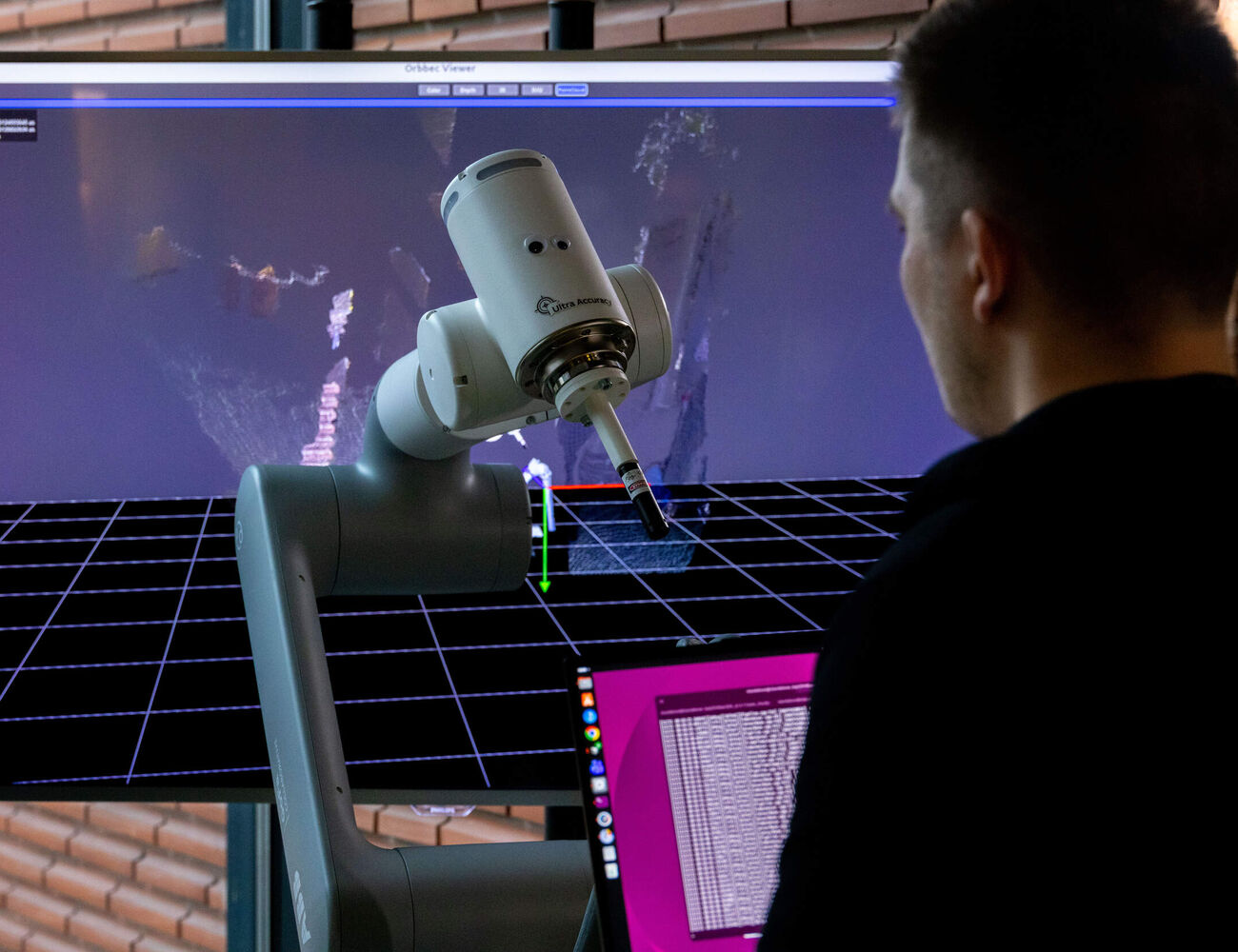
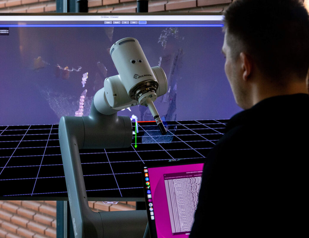
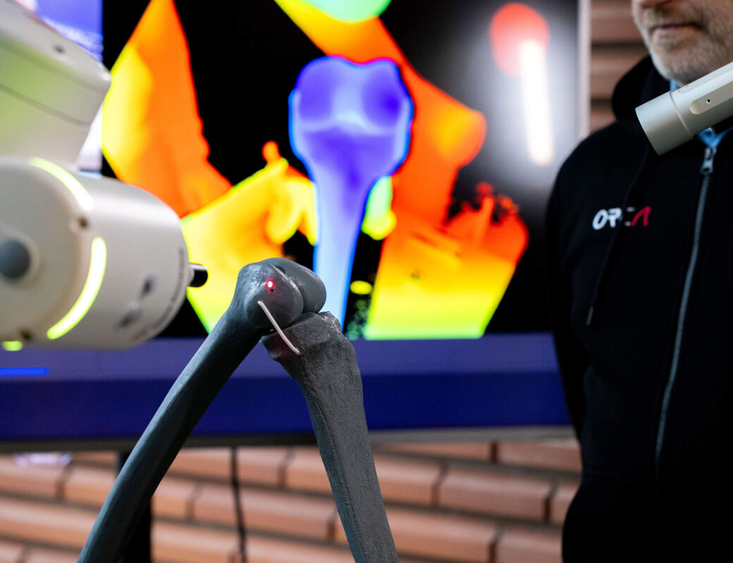
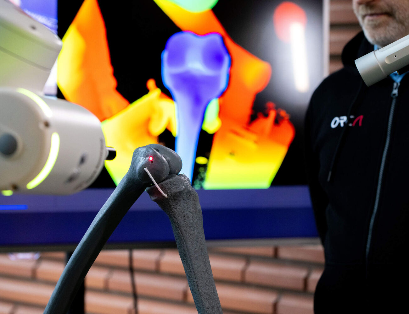
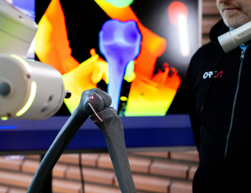
There’s also an essential difference in how the systems work. The current system tracks the reflective spheres and infers the bone’s position, whereas a 3D camera measures the bone directly. The machine vision application recognises the bone in the point cloud produced by the camera and determines its position. ‘Our navigation system can distinguish bone from other tissue,’ says Pitkänen.
Already accurate enough
The Aalto researchers have optimised the camera to make the system accurate enough for clinical use. They filed their first patent applications for this in October 2026.
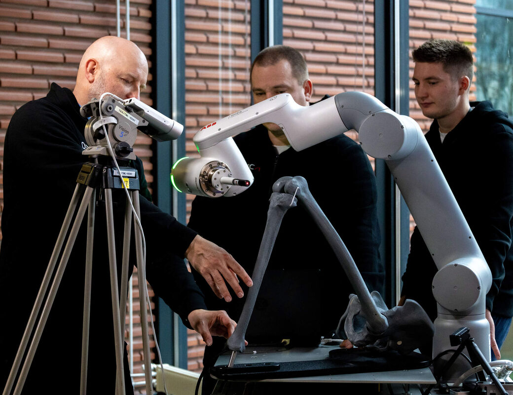
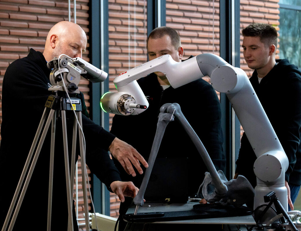
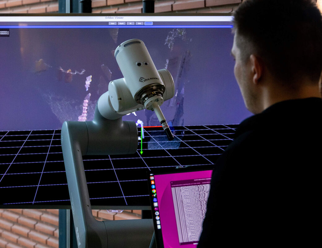
In their tests, the team connected their system to a GoFa robot manufactured by ABB. Similar robots are commonly used in the electronics industry, where high precision is required. When Pitkänen attached a laser to the robotic arm, it pointed at exactly the same spot on a plastic bone every time, even if the spot wasn’t in the camera’s view. When the bone was moved, the laser continued tracking the point.
‘We can see a certain part of the bone, and then we know where the whole bone is. We don’t need to see the spot where the drilling will take place,’ he explains.

The researchers have also tested their system on animal bones. They have a whole pig’s leg in the freezer which the food company Atria supplied for their research.
‘In the next stage, we’ll test the system on cadavers donated for medical research, in collaboration with doctors. We also intend to take our navigation system into the operating theatre to observe joint replacement operations,’ says Pitkänen.
Helping hospitals
The idea of using a 3D camera doesn’t come solely from Pitkänen’s experience with surgical robotics. The team members also recognised the need for it when they took part in the Biodesign Finland programme. The programme, developed at Stanford University, brings together multidisciplinary teams to identify problems in healthcare and devise solutions that could be commercialised. Aalto and HUS host Biodesign Finland together each year.
‘We spent a few months walking around HUS’s Siltasairaala hospital looking for problems and coming up with solutions,’ Pitkänen says.
After selecting their research topic, the group established the ORCA project and secured Business Finland Research to Business funding, which aims to help prepare research results for commercialisation.
‘We’re currently exploring different commercialisation options. One is licensing the technology, and another is setting up a start-up.’
Pitkänen believes the market is enormous, because the navigation system developed at Aalto could be connected to the control system of any robot. It could also serve as an extra set of eyes in joint replacement operations where the surgeon isn’t using a robot.
‘An easy-to-use system could make computer-assisted navigation an attractive option for hospitals where buying an expensive robot does not make sense. It might cost a tenth of the price of a robot,’ Pitkänen says.
Once the 3D camera has proven its capabilities in knee replacement surgery, the research group will move on to other types of joint replacement and bone surgery in general, such as spinal and brain surgery. ‘All of them use reflective markers. We want to replace those with a 3D camera.’
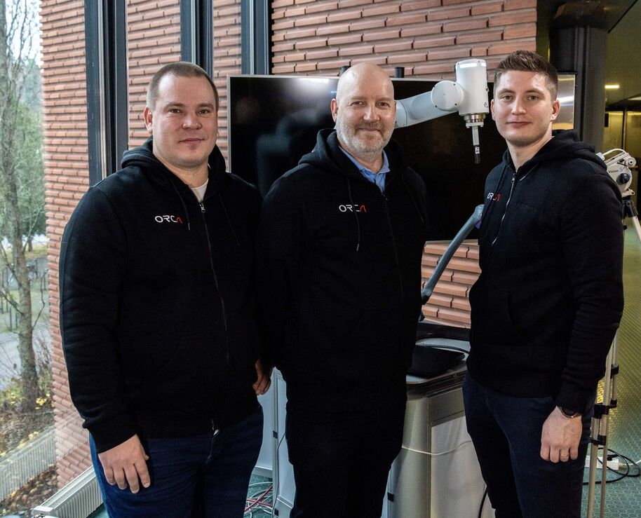
Machine vision with simplicity and precision
Why hasn’t machine vision-based navigation become established in joint replacement surgery? The Aalto ORCA research group, which is developing the technology, has a few explanations.
‘One reason may be that computing power is only now reaching a sufficient level. A lot of computation is needed to track the tens of thousands of points produced by a 3D camera,’ says Sauli Lindroos, who focuses on biomedical imaging
Even with enough computing power, it takes the right methods to make it work. Lindroos explains that machine vision algorithms – computational methods – have developed to the point where objects can be recognised and located more quickly and accurately than just a few years ago. This makes it possible, for example, to distinguish bone from the surrounding tissue.
It might be tempting to talk about artificial intelligence in this context, but the research group is wary of the term. That could bring to mind generative AI, which can produce different answers from the same input. ORCA’s aim is the opposite.
‘We have chosen established algorithms that are predictable and repeatable. When given the same input, they always produce the same result,’ says Taneli Tikkakoski, whose background is in bioinformatics.
The result is machine vision that is simple and precise. It doesn’t think for the surgeon or learn on the fly. It simply sees and measures accurately.
Text: Marianna Salin
Photos: Mikko Raskinen
Aalto at Slush 2026: These solutions are revolutionising the insulation market, joint replacement surgery and the utilisation of waste heat
Aalto University will take part in the startup event Slush by showcasing 12 new innovations that help solve sustainability challenges and strengthen economic growth.

Read more news

Finding beauty in grey November
Dreary autumn skies getting you down? Aesthetics researcher Sanna Lehtinen says there’s infinite beauty in the shades of grey––if only we take the time to look.
Aalto and Unite! develop new teaching modules through international collaboration
Aalto joined three other Unite! partner universities to develop 11 shared teaching modules in engineering, cybersecurity, robotics and ethics, bringing new expertise into teaching across the alliance.
999 Ways of Shaping Nature: an exhibition by visiting researcher Cristiana Macedo
Visiting researcher Cristiana Macedo shows 999 Ways of Shaping Nature, made with soils from Portugal and Finland, in the F Lobby at Väre, 16–22 November 2026.