New scanner can improve the detection of cancer tissue and brain disease diagnoses
Researchers aim to have the technology ready for hospital use and commercialisation by the end of next year.
Quantum-mechanical phenomena such as superconductivity and nuclear magnetic resonance allow probing the human brain and detecting its function using magnetic fields. Developing the MEG-MRI methodology and instrumentation involves a variety of topics such as physics, electronics, neuroscience, prototyping, signal processing, mathematics and programming (most notably Python). MEG-MRI is a key part in the BREAKBEN EU Consortium aiming at dramatic improvements in electromagnetic neuroimaging.
A relatively new technique, where the nuclear magnetic resonance (NMR) is measured at kHz frequencies using ultra-sensitive magnetic field sensors, typically based on superconducting quantum-interference devices (SQUIDs) and operating at a 4.2 K temperature. ULF MRI provides a completely silent and safe method for acquiring 3D images, for instance, of the human head.
CDI maps the electric current density flowing in a volume, such as in living tissues in the human head. ULF MRI offers unique possibilities in current-density imaging, because with ultra-low-noise electronics developed in the MEG-MRI group, all the applied magnetic fields can be rapidly switched on and off during the imaging sequence. The group has also published the first CDI pulse sequences and simulations based on this technique, and we proceed to develop the technique in practice. Current-density imaging can provide valuable information for improving the accuracy of localization of electrical brain activity.
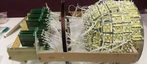
MEG is the measurement of the magnetic field generated by electric currents in neuronal activity. When a large number of highly sensitive magnetic field sensors is used, one obtains information about the location of the brain activity. A great deal of MEG expertise is concentrated in the Helsinki area. The combination of MEG with ULF MRI and CDI can signifantly improve the workflow and accuracy of localizing brain activity, allowing also experimentation with new techniques.
For more information, contact the group leader Cornelis Koos Zevenhoven at [email protected]
Researchers aim to have the technology ready for hospital use and commercialisation by the end of next year.
The EU-funded BREAKBEN project promises better and more detailed brain scans
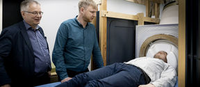
The aim of the project is to develop an instrument combining magnetic resonance imaging (MRI) and magnetoencephalography (MEG).