Measuring forces of living cells and microorganisms

Forces that are exerted by a living cell or a microorganism are tiny and often not larger than a few nanonewtons. For comparison, one nanonewton is the weight of one part in a billion of a typical chocolate bar. Yet, for biological cells and microbes, these forces are enough to allow cells to stick to a surface or microbes to propel themselves towards nutrients. Scientists from Finland and Germany now present a highly adaptable technique, called ‘micropipette force sensors’, to precisely measure the forces exerted by a wide range of micron-sized organisms. This novel method has now been published in the journal Nature Protocols.
To stay alive and proliferate, a biological cell needs to be able to successfully adapt to its environmental conditions. The ability to do so involves physical principles and mechanical forces: cells may attach themselves to surfaces and other cells to eventually form a biofilm, a structure which protects the community of cells from external attack. Many microorganisms can actively move themselves, by crawling on a surface or swimming in liquid, for example, towards a source of nutrients. In order to advance our fundamental understanding of how microbes can move themselves, it’s important for us to be able to measure the mechanical forces associated with their movement.
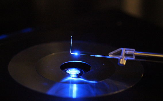
The development of micropipette force sensors to measure forces of living cells and microorganisms is described in a joint work by Dr. Matilda Backholm and Dr. Oliver Bäumchen. 'The working principle of the micropipette force sensor technique is beautifully simple: by optically observing the deflection of a calibrated micropipette, the forces acting on the pipette can be directly measured,' says Matilda Backholm, researcher at the Department of Applied Physics of Aalto University in Finland.
A micropipette is a hollow glass needle featuring a thickness of about the diameter of a human hair or even smaller. One of the most remarkable advantages of this technique is the fact that it can be applied to a large variety of biological systems, ranging from a single cell to a millimeter-size microorganism. 'We exemplified the versatility of our method using two model systems from microbiology, but certainly the technique can and will be applied to other biological systems in the future,' says Oliver Bäumchen, research group leader at the Max Planck Institute of Dynamics and Self-Organization in Göttingen, Germany.
'The idea behind the technique is to combine the advantages of several established biophysical techniques: we use a micropipette to grab a living cell, in the exact same way as it is done in in-vitro fertilization, and study the mechanical forces by measuring the pipette’s deflection using the measurement principles underlying atomic force microscopy – a standard measurement technique in physics,' says Bäumchen. Dr. Backholm points out another major advantage: “In contrast to other force measurement methods, we detect the deflection of our highly sensitive micropipette simply by observing it with a state-of-the-art microscope. This allows us to inspect the shape and motion of the microorganism with high optical resolution, while we are measuring the forces simultaneously.” During all of this, the cell or microorganism is fully intact and alive, which allows for testing its reaction to drugs as well as nutrients, temperature and other environmental factors. 'The force resolution is really remarkable. With our recent technological advancements, we successfully managed to detect forces down to about ten piconewtons, which is almost as good as an atomic force microscope,' adds Dr. Bäumchen.
The researchers expect that their method will be applied in other research labs in the future to tackle a plethora of important biophysical questions, aiming at better understanding biological functions of cells and microorganisms, as well as their underlying physical principles. Dr. Backholm points out that these research avenues may indeed advance biomedical and biotechnological applications: 'The micropipette force sensor technique might help to identify drugs for fighting infectious diseases and inhibiting the formation of biofilms on medical implants, just to name a few examples where this novel approach might make a significant impact.'
Original Publication
Micropipette force sensors for in vivo force measurements on single cells and multicellular microorganisms
Matilda Backholm & Oliver Bäumchen, Nature Protocols, 28. January 2019,
https://doi.org/10.1038/s41596-018-0110-x
Additional images and data in the video from Dr Rafael Schulman
Read more news

Apply Now: Unite! Visiting Professorships at TU Graz
TU Graz, Austria, invites experienced postdoctoral researchers to apply for two fully funded visiting professorships. The deadline for expressions of interest is 20 February 2026, and the positions will begin on 1 October 2026.
Hanaholmen’s 50th anniversary exhibition lives on online – making the history of Finnish–Swedish cooperation accessible worldwide
MeMo Institute at Aalto University has produced a virtual 3D version of the anniversary exhibition of Hanaholmen.Soil Laboratory Exhibition – Exploring the Dialogue Between Human and the Earth in Utsjoki
Soil Laboratory explores the relationship between humans and the earth as a living landscape through ceramic practices in Utsjoki.


