


Scientists use bacteria as micro-3D printers
A team at Aalto University has used bacteria to produce intricately designed three-dimensional objects made of nanocellulose. With their technique, the researchers are able to guide the growth of bacterial colonies through the use of strongly water repellent – or superhydrophobic – surfaces. The objects show tremendous potential for medical use, including supporting tissue regeneration or as scaffolds to replace damaged organs. The results have been published in the journal ACS Nano.
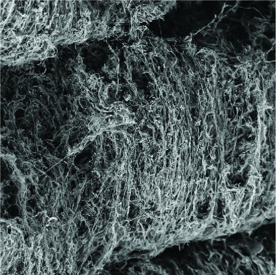
Unlike fibrous objects made through current 3D printing methods, the new technique allows fibres, with a diameter a thousand times thinner than a human hair, to be aligned in any orientation, even across layers, and various gradients of thickness and topography, opening up new possibilities for application in tissue regeneration. These kinds of physical characteristics are crucial for support materials in the growth and regeneration of certain types of tissues found in muscles as well as in the brain.
‘It’s like having billions of tiny 3D printers that fit inside a bottle,’ explains Luiz Greca, a doctoral student at Aalto University. ‘We can think of the bacteria as natural microrobots that take the building blocks provided to them and, with the right input, create complex shapes and structures.’
Once in a superhydrophobic mould with water and nutrients — sugar, proteins and air — the aerobic bacteria produce nanocellulose. The superhydrophobic surface essentially traps a thin layer of air, which invites the bacteria to create a fibrous biofilm replicating the surface and shape of the mould. With time, the biofilm grows thicker and the objects become stronger.
Using the technique, the team has created 3D objects with pre-designed features, measuring from one-tenth the diameter of a single hair all the way up to 15-20 centimetres. The nano-sized fibres do not cause adverse reactions when placed in contact to human tissues. The method could also be used to grow realistic models of organs for training surgeons or improving the accuracy of in-vitro testing.
Luiz Greca, doctoral studentIt’s like having billions of tiny 3D printers that fit inside a bottle.
‘It’s really exciting to expand this area of biofabrication that takes advantage of strong cellulose nanofibres and the networks they form. We’re exploring applications for age-related tissue degeneration, with this method being a step forward in this and other directions,’ says research group leader Professor Orlando Rojas. He adds that the strain of bacteria used by the team, Komagataeibacter medellinensis, was discovered in a local market in the city of Medellin, Colombia, by previous collaborators from Universidad Pontificia Bolivariana.
In both nature and engineering, superhydrophobic surfaces are designed to minimise the adhesion of dust particles as well as microorganisms. This work is expected to open new possibilities for using superhydrophobic surfaces to precisely produce naturally manufactured materials.
As the bacteria can be removed or left in the final material, the 3D objects can also evolve as a living organism over time. The findings provide an important step towards harnessing full control over bacterially fabricated materials.
‘Our research really shows the need to understand both the fine details of bacteria interaction at interfaces and their ability to make sustainable materials. We hope that these results will also inspire scientists working on both bacteria-repelling surfaces and those making materials from bacteria,’ says Dr. Blaise Tardy.
Publication:
Greca, L. G., Rafiee, M., Karakoç, A., Lehtonen, J., Mattos, B. D., Tardy, B. L., & Rojas, O. J. (2020). Guiding Bacterial Activity for Biofabrication of Complex Materials via Controlled Wetting of Superhydrophobic Surfaces. ACS Nano. https://doi.org/10.1021/acsnano.0c03999
More information
Read more news

The company established by three anthropologists relied on strong research expertise
Etnografinen tutkimustoimisto Kenno Oy was established in 2014 when its three founders were finalising their master's degree studies at the University of Helsinki and wanted to create an interesting job for themselves.
Bachelor's Students Successfully Complete Research Internship
Three bachelor's students have successfully completed their research internships.
From Materials to Smart Textiles: Benjamin Begins a New Chapter at Aalto
Ping-Chun Chen is a researcher from National Tsing Hua University, Taiwan.