


Story of magnetic resonance imaging device tells of courage and determination

The young Master of Science and Engineering Raimo Sepponen read through scientific articles one after another. He was interested in new methods which could be used for diagnosing internal haemorrhages without requiring surgical measures. By chance, one discovery caught his attention. Nuclear magnetic resonance imaging (NMR) was still a new research method at the end of the 1970s. It could be used to view the water distribution in body tissue. Sepponen wandered if this could be a solution, and decided to find out more by heading to London for the first international conference on the topic.
Raimo Sepponen was working at that time for Instrumentarium Ltd., which made the bold long-term decision to shift from importing to developing new export products. An international survey was carried out by the company to find potential problems that they could seek to solve, and a non-invasive diagnosis method and device for internal haemorrhages was chosen as one development project. Before the development of present-day magnetic resonance imaging devices, internal haemorrhages could generally only be diagnosed through surgical operations.
‘For example, diagnosis of a haemorrhage in the abdominal cavity was generally done using a physiological saline solution. When the solution was taken out of the abdominal cavity, the bloodiness of the liquid was used to decide whether the patient had a internal haemorrhage or not,’ Sepponen remembers. Haemorrhages within organs or within the head, however, could not be detected using this method. Precise information could not be obtained, and so the patient instead had to be operated on in order to see where the haemorrhage was.
The company and the researchers were certain that this was a problem that was worth solving, even though not even all doctors were in agreement on the need for such an invention. A doctor at the Töölö Hospital Trauma Centre stated that a device for observing internal haemorrhages was not needed because ‘we simply make a small hole, peak in and see if there is blood or not.’
Professor Emeritus Raimo SepponenWith the current investment level and funding model, it’s doubtful we can get things moving.
Into the unknown
There was no guarantees that the technology would work. At the start of the project, it wasn’t known what a haemorrhage would look like in the images. ‘We imagined that the blood would accumulate and form a lump in one place, whereas it can actually be a thin film between internal organs.’
Based on research carried out in other research institutes, it was possible to conclude that some kind of images would be obtained. It took a long time, however, to obtain each image. At the end of the 1970s, taking one image took four hours. Compared to that, the 10-minute imaging time obtained by the start of the 1980s was already very short.
Building the first prototype of the device was a demanding but ultimately rewarding project. The expertise and working environment of the Helsinki University of Technology’s Low Temperature Laboratory enabled the project to succeed. When in Autumn 1981, imprecise brain images comparable to present-day brain images were obtained for the first time, the researchers went mad with delight. Bit by bit, they accumulated more expertise and information about how to further develop the device and obtain more precise images. In the end, the device was already in hospital use just a year after the first image was successfully obtained. The device was among the first magnetic resonance imaging devices to be used in a hospital anywhere in the world, and it was used to scan over 3500 patients.
In hindsight, Sepponen is amazed at the strong support given by the company and the management’s farsightedness. In Sepponen’s opinion, the same kind of boldness and commitment to technological development cannot be seen nowadays, even though this is just what is needed if the goal is to make health technology a significant export sector for Finland.
‘Health technology exports are currently at €2 billion. By 2030, this should be between €10–20 billion, if the goal is to make health technology exports a significant economic driver’, Sepponen says.
‘With the current investment level and funding model, it’s doubtful we can get things moving. In addition to funding, we need both ideas and companies ready for change.’
The train has left already
The future has a lot in common with the past. Sepponen shares the story of the magnetic resonance imaging device because it can be used to learn for the future. The company made a conscious decision to set out to develop something totally new. With the help of an international jury, a problem affecting a sufficiently large number of people was chosen and then the search for a solution began. Knowing that they had secure long-term funding, the researchers were able to focus on the research.
Sepponen emphasises that expertise must be developed to the highest level so that we are ready at the right moment to offer customers the solution they need. He believes that Finland has significant business opportunities, particularly in the areas of rehabilitation and disease prevention. Expertise and products must be developed now so that we are ready when the opportunity for success is upon us.
‘We need to be keeping watch, as an ice fisher does by the edge of the ice hole, waiting for the right moment and offering as tempting a bait as possible. It’s too late to start up the engine once the markets are livening up, because the competitors are then already much further ahead.’

The Finnish Society for Medical Physics and Medical Engineering made Professor Emeritus Raimo Sepponen an honorary member at its 50th anniversary celebrations in October 2018. The Radiological Society of Finland awarded the Carl Wegelius medal to Professor Emeritus Raimo Sepponen as a recognition of his work for the development of magnetic resonance imaging. Professor Sepponen gave the Carl Wegelius lecture on the topic ‘Health technology as a Finnish economic pillar’ at the opening ceremony of the Radiation Protection Days in November 2018.
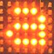
A magnetic resonance imaging device is just one example of a technology developed through the need to solve a particular problem. As examples of other health technologies developed in Finland, Sepponen mentions the dental x-ray machine, the anaesthesia monitoring device and the mammography device. The first images were still very imprecise compared to the kind of images that can be obtained nowadays (see the main picture).
Professor Emeritus Raimo Sepponen has worked with health technology for his whole career:
- Researching impedance cardiography, cover dilution methods and cardiac perfusion modelling at HYKS’s cardiology department (1975)
- Master’s thesis on indirect blood pressure measurement for Ollituote Oy (name later changed to Kone Instruments Oy) (1974)
- Licentiate thesis on indirect blood pressure measurement using impedance methods (1979)
- Development of continuous flow analyser to be used in Instrumentarium/Datex Hospital Laboratory (1976–1977)
- Development of point-of-care analyser for heart attacks (1976)
- Development of health centre photometry (1977)
- Non-invasive diagnosis method and device for internal haemorrhages => magnetic resonance imaging (1977–1995)
- HUT: Ballistocardiography, incl. bathroom scales for measuring heart rate (1994–
- A health chair which measures a number of vital signals and identifies trends (weight, ECG, chest impedance, pulse oximetry and blood pressure), for use in assisted living accommodation, health kiosks and other such locations (1996–
- The Ballistocardiography research gave birth to the company Finnsor Oy, which is led by Lasse Leppäkorpi and which later become Beddit Oy, now owned by Apple.
- Smart floor: the ‘ELSI sensor’ for recognising falls and a need for help during the night, used in assisted living accommodation and other such locations. Currently a product of the company Mari Care (2000–2009)
- Smart nappy for measuring the moisture content of a chronic invalid’s nappy. This is now applied by the company Vigilan Oy to the measurement of moisture in building structures. (1997–2004)
Text: Riikka Hopiavaara
Read more news

Catalysis in a new light: Microscale interactions could enhance clean energy technologies
A new study provides a more detailed view of how catalysts function during chemical reactions. The discovery could help develop more efficient materials for applications such as green hydrogen production and a more sustainable chemical industry.
Physics Days 2026 gathered Finnish physicists to Aalto
The 2026 edition of the annual conference featured talks on moiré matter, women in physics and paper cuts.
Annual review looked back on the past year
The annual review of the School of Arts, Design and Architecture provided a comprehensive overview of the past year. Members of the community were also awarded in the event.