Artificial intelligence to assist the brain
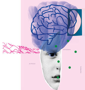
The human brain consists of some 86 billion neurons, nerve cells that process and convey information through electrical nerve impulses.
That’s why measuring neural electrical activity is often the best way to study the brain, says Hanna Renvall. She is Aalto University and HUS Helsinki University Hospital Assistant Professor in Translational Brain Imaging and heads the HUS BioMag Laboratory.
Electroencephalography, or EEG, is the most used brain imaging technique in the world. Renvall's favourite, however, is magnetoencephalography or MEG, which measures the magnetic fields generated by the brain’s electrical activity.
MEG signals are easier to interpret than EEG because the skull and other tissues don’t distort magnetic fields as much. This is precisely what makes the technique so great, Renvall explains.
‘MEG can locate the active part of the brain with much greater accuracy, at times achieving millimetre-scale precision.’
An MEG device looks a lot like bonnet hairdryers found in hair salons. The SQUID sensors that perform the measurements are concealed and effectively insulated inside the bonnet because they only function at truly freezing temperatures, close to absolute zero.
The world's first whole-head MEG device was built by a company that emerged from Helsinki University of Technology’s Low Temperature Laboratory – and is now the leading equipment manufacturer in this field.
MEG plays a major role in the European Union’s new AI-Mind project, whose Finnish contributors are Aalto and HUS. The goal of the €14-million project is to learn ways to identify those patients, whose dementia could be delayed or even prevented.
For this to happen, neuroscience and neurotechnology need help from artificial intelligence experts.
Fingerprinting the brain
Dementia is a broad-reaching neural function disorder that significantly erodes the sufferer’s ability to cope with everyday life. Some 10 million people are afflicted in Europe, and as the population ages this number is growing. The most common illness that causes dementia is Alzheimer’s disease, which is diagnosed in 70–80% of dementia patients.
Researchers believe that communication between neurons begins to deteriorate well before the initial clinical symptoms of dementia present themselves. This can be seen in MEG data—if you know what to look for.
MEG is at its strongest when measuring the brain’s response to stimuli like speech and touch that occur at specific moments and are repetitive.
Interpreting resting-state measurements is considerably more complex.
That’s why the AI-Mind project uses a tool referred to as the fingerprint of the brain. It was created when Renvall and Professor Riitta Salmelin and her colleagues began to investigate whether MEG measurements could detect a person’s genotype. More than 100 sibling pairs took part in the study that sat subjects in an MEG, first for a couple of minutes with their eyes closed and then for a couple of minutes with their eyes open. They also submitted blood samples for a simple genetic analysis.
When researchers compared the graphs and genetic markers, they noticed that, even though there was substantial variance between individuals, siblings’ graphs were similar.
Next, Aalto University Artificial Intelligence Professor Samuel Kaski’sgroup tested whether a computer could learn to identify graph sections that were as similar as possible between siblings while also being maximally different when compared to other test subjects.
The machine did it—and more, surprisingly.
‘It learned to distinguish the individual perfectly based on just the graphs, irrespective of whether the imaging had been performed with the test subject’s eyes open or closed,’ Hanna Renvall says.
‘For humans, graphs taken with eyes closed or open look very different, but the machine could identify their individual features. We’re extremely excited about this brain fingerprinting and are now thinking about how we could teach the machine to recognise neural network deterioration in a similar manner.’
Risk screening in one week
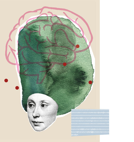
A large share of dementia patients are diagnosed only after the disorder has already progressed, which explains why treatments tend to focus on managing late-stage symptoms.
Earlier research has, however, demonstrated that many patients experience cognitive deterioration, such as memory and thought disorders, for years before their diagnosis.
One objective of the AI-Mind project is to learn ways to screen individuals with a significantly higher risk of developing memory disorders in the next few years from the larger group of those suffering from mild cognitive deterioration.
Researchers plan to image 1,000 people from around Europe who are deemed at risk of developing memory disorders and analyse how their neural signals differ from people free from cognitive deterioration. AI will then couple their brain imaging data with cognitive test results and genetic biomarkers.
Researchers believe this method could identify a heightened dementia risk in as little as a week.
‘If people know about their risk in time, it can have a dramatic motivating effect,’ says Renvall, who has years of experience of treating patients as a neurologist.
Lifestyle changes like a healthier diet, exercise, treating cardiovascular diseases and cognitive rehabilitation can significantly slow the progression of memory disorders.
Better managing risk factors can give the patient many more good years, which is tremendously meaningful for individuals, their loved ones and society, as well, Renvall says.
Identifying at-risk individuals will also be key when the first drugs that slow disease progression come on the market, perhaps in the next few years. Renvall says it will be a momentous event, as the medicinal treatment of memory disorders has not seen any substantial progress in the last two decades.
The new pharmaceuticals will not suit everybody, however.
‘These drugs are quite powerful, as are their side effects—that’s why we need to identify the people who can benefit from them the most,’ Renvall emphasises.
Zapping the brain
Brain activity involves electric currents, which generate magnetic fields that can be measured from outside the skull.
The process also works in the other direction, the principle on which transcranial magnetic stimulation (TMS) is based. In TMS treatments, a coil is placed on the head to produce a powerful magnetic field that reaches the brain through skin and bone, without losing strength. The magnetic field pulse causes a short, weak electric field in the brain that affects neuron activity.
It sounds wild, but it’s completely safe, says Professor of Applied Physics Risto Ilmoniemi, who has been developing and using TMS for decades.
‘The strength of the electric field is comparable to the brain’s own electric fields. The patient feels the stimulation, which is delivered in pulses, as light taps on their skin.’
Magnetic stimulation is used to treat severe depression and neuropathic pain. At least 200 million people around the world suffer from severe depression, while neuropathic pain is prevalent among spinal injury patients, diabetics and multiple sclerosis sufferers. Pharmaceuticals provide adequate relief to only half of all depression patients; this share is just 30% in the case of neuropathic pain sufferers.
How frequently pulses are given is based on the illness being treated. For depression, inter-neuron communication is stimulated with high-frequency pulse series, while less frequent pulses calm patient’s neurons for neuropathic pain relief.
Stimulation is administered to the part of the brain where, according to the latest medical science, the neurons tied to the illness being treated are located.
About half of treated patients receive significant relief from magnetic stimulation. Ilmoniemi believes this could be much higher—with more coils and the help of algorithms.
One-note clanger to concert virtuoso
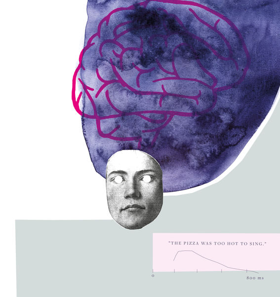
In 2018, the ConnectToBrain research project headed by Ilmoniemi was granted €10 million in European Research Council Synergy funding, the first time that synergy funds were awarded to a project steered by a Finnish university. Top experts in the field from Germany and Italy are also involved.
The goal of the project is to radically improve magnetic stimulation in two ways: by building a magnetic stimulation device with up to 50 coils and by developing algorithms to automatically control the stimulation in real time, based on EEG feedback.
Ilmoniemi looks to the world of music for a comparison.
‘The difference between the new technology and the old is analogous to a concert pianist playing two-handed, continuously fine-tuning their performance based on what they hear, rather than hitting a single key while wearing hearing protection.’
Researchers have already used a two-coil device to demonstrate that an algorithm can steer stimulation in the right direction ten times faster than even the most experienced expert. This is just the beginning.
A five-coil device completed last year covers an area of ten square centimetres of cortex at a time. A 50-coil system would cover both cerebral hemispheres.
Building this kind of device involves many technical challenges. Getting all these coils to fit around the head is no easy task, nor is safely producing the strong currents required.
Even once these issues are resolved, the hardest question remains: how can we treat the brain in the best possible way?
‘What kind of information does the algorithm need? What data should instruct its learning? It is an enormous challenge for us and our collaborators,’ Ilmoniemi says thoughtfully.
The project aims to build one magnetic stimulation device for Aalto, another for the University of Tübingen in Germany and a third for the University of Chieti-Pescara in Italy. The researchers hope that, in the future, there will be thousands of such devices in operation around the world.
‘The more patient data is accumulated, the better the algorithms can learn and the more effective the treatments will become.’
Hanna RenvallThe machine learned to distinguish the individual perfectly based on just the graphs.
Quantum optics sensors could revolutionise how we read neural signals
Professor Lauri Parkkonen’s working group is developing a new kind of MEG device that adapts to the head size and shape and utilises sensors based on quantum optics. Unlike the SQUID sensors currently employed in MEG, they do not need to be encased in a thick layer of insulation, enabling measurements to be taken closer to the scalp surface. This makes it easier to perform precise measurements on children and babies especially.
The work has progressed at a brisk pace and yielded promising results: measurements made with optical sensors are already approaching the spatial accuracy of measurements made inside the cranium.
Parkkonen believes that a MEG system based on optical sensors could also be somewhat cheaper and more compact and thus easier to place than traditional devices; such a MEG system could utilize a ‘person-sized’ magnetic shield instead of a large shielded room as the conventional MEG systems do.
‘This would bring it into reach of more researchers and hospitals.’
This article has been published in the Aalto University Magazine issue 30 (issuu.com), April 2022.
Donate to develop
Aalto University's public core funding from the Ministry of Education and Culture has decreased in real terms by more than one-third compared to 2010, when the university began its operations. As a society, we cannot afford to cut back on education and research.
To ensure groundbreaking research and bold, persistent innovators in the future, we must invest in both long-term fundamental research and targeted innovation efforts. Donate and you’ll invest in a future you can be proud of.
School of Science
School of Science
Translating research findings into clinical patient care is a long process that builds on previous scientific advancements. Medical research and technological developments are advanced at the Department of Neuroscience and Biomedical Engineering, in the School of Science.
Read more news
Soil Laboratory Exhibition – Exploring the Dialogue Between Human and the Earth in Utsjoki
Soil Laboratory explores the relationship between humans and the earth as a living landscape through ceramic practices in Utsjoki.
The Finnish Cultural Foundation awarded grants for science and art
A total of 15 individuals or groups from Aalto University received grants
Environmental Structure of the Year 2025 Award goes to Kalasatama-Pasila tramway
The award is given in recognition of meritorious design and implementation of the built environment. Experts from Aalto University developed sustainability solutions for the project.


