HRMEG

High-resolution MEG: towards non-invasive corticography
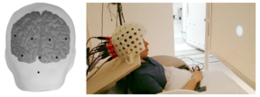
To date, neuroimaging has provided a wealth of information on how the human brain works in health and disease. With functional magnetic resonance imaging (fMRI), we can obtain spatially precise information about long-lasting brain activations whereas electro- and magnetoencephalography (EEG/MEG) can track transient cortical responses at millisecond resolution. However, none of these methods excel in time-resolved detection of sustained cortical activations, which are typically reflected as bursts of gamma-range (30–150 Hz) oscillations, frequently present in invasive recordings in patients. Although we have recently demonstrated that in exceptional situations MEG can detect even single gamma responses, their signal-to-noise ratio is usually prohibitively low, largely due to the substantial distance (4–5 cm) between cortex and sensors. In this HRMEG project, we exploit recent advances in a novel magnetic sensor technology—optically-pumped magnetometeres (OPM)—to construct a new kind of MEG system that allows capturing cerebral magnetic fields within millimetres from the scalp. Our simulations show that this proximity leads up to a 5-fold increase in the signal amplitude and an order-of-magnitude improvement of spatial resolution compared to conventional MEG based on superconductive SQUID sensors. Therefore, a high-resolution MEG system based on OPMs should enable non-invasive recordings of cortical activity at unprecedented sensitivity and detail level, which we propose to capitalize on by characterizing cortical responses, particularly gamma oscillations, during complex cognitive tasks. Additionally, since OPMs can recover within milliseconds from fields of several tesla, we will also seek to combine transcranial magnetic stimulation (TMS) with MEG, leveraging the reciprocity of TMS and MEG and thus allowing better-than-ever characterization of TMS-evoked responses.
In summary, the HRMEG project aims at providing a new kind of time-resolved neuroimaging method, which can target smaller functional units of the human cortex than currently possible with any non-invasive technique. In the future, the approach could also open new avenues in developmental neuroscience as the new sensor array could be optimally fitted to infants and children, unlike the sensor arrays of current MEG systems. Similarly, improvements could be expected in clinical diagnostics due to the increased sensitivity to sustained high-frequency activity. In addition, HRMEG could be useful in screening patients who would benefit from an implanted BCI system.
The HRMEG project is funded by European Research Council (ERC) Starting Grant to prof. Lauri Parkkonen, who is the principal investigator of this project.
Current state of the project
So far the project has focused on the development of the measurement system and on its validation to perform measurements of human brain function. To this end, we have
- designed and constructed the OPM sensor array with over 20 sensors that can be freely positioned on the subject's head,
- created a subsystem for suppressing both static and dynamic ambient magnetic interference,
- built a data acquisition subsystem that allows vizualizing and pre-processing the signals,
- developed and tested means for co-registering sensor positions to structural magnetic resonance images of the brain in order to enable source estimation, and
- performed human brain measurements with the system to verify that the whole approach works as intended.
The above progress is documented in detail in the publications listed below. Currently we are expanding the system to include more sensors and applying it to record brain responses to a variety of sensory stimuli.
Project team

Peer-reviewed publications:
- Iivanainen, J., Borna, A., Zetter, R., Carter, T.R., Stephen, J.M., McKay, J., Parkkonen, L., Taulu, S., Schwindt, P.D.D., 2022. Calibration and Localization of Optically Pumped Magnetometers Using Electromagnetic Coils. Sensors (Basel) 22, 3059. https://doi.org/10.3390/s22083059
- Iivanainen, J., Mäkinen, A.J., Zetter, R., Stenroos, M., Ilmoniemi, R.J., Parkkonen, L., 2021a. Spatial sampling of MEG and EEG based on generalized spatial-frequency analysis and optimal design. NeuroImage 245, 118747. https://doi.org/10.1016/j.neuroimage.2021.118747
- Iivanainen, J., Mäkinen, A.J., Zetter, R., Zevenhoven, K.C.J., Ilmoniemi, R.J., Parkkonen, L., 2021b. A general method for computing thermal magnetic noise arising from thin conducting objects. Journal of Applied Physics 130, 043901. https://doi.org/10.1063/5.0050371
- Helle, L., Nenonen, J., Larson, E., Simola, J., Parkkonen, L., Taulu, S., 2021c. Extended Signal-Space Separation Method for Improved Interference Suppression in MEG. IEEE Trans Biomed Eng 68, 2211–2221. https://doi.org/10.1109/TBME.2020.3040373
- Iivanainen, J., Zetter, R., Parkkonen, L., 2020a. Potential of on-scalp MEG: Robust detection of human visual gamma-band responses. Human Brain Mapping 41, 150–161. https://doi.org/10.1002/hbm.24795
- Mäkinen, A.J., Zetter, R., Iivanainen, J., Zevenhoven, K.C.J., Parkkonen, L., Ilmoniemi, R.J., 2020b. Magnetic-field modeling with surface currents. Part I. Physical and computational principles of bfieldtools. Journal of Applied Physics 128, 063906. https://doi.org/10.1063/5.0016090
- Zetter, R., J. Mäkinen, A., Iivanainen, J., Zevenhoven, K.C.J., Ilmoniemi, R.J., Parkkonen, L., 2020c. Magnetic field modeling with surface currents. Part II. Implementation and usage of bfieldtools. Journal of Applied Physics 128, 063905. https://doi.org/10.1063/5.0016087
- Iivanainen, J., Zetter, R., Grön, M., Hakkarainen, K., Parkkonen, L., 2019a. On-scalp MEG system utilizing an actively shielded array of optically-pumped magnetometers. NeuroImage 194, 244–258. https://doi.org/10.1016/j.neuroimage.2019.03.022
- Zetter, R., Iivanainen, J., Parkkonen, L., 2019b. Optical Co-registration of MRI and On-scalp MEG. Scientific Reports 9, 5490. https://doi.org/10.1038/s41598-019-41763-4
- Zubarev, I., Zetter, R., Halme, H.-L., Parkkonen, L., 2019c. Adaptive neural network classifier for decoding MEG signals. NeuroImage. https://doi.org/10.1016/j.neuroimage.2019.04.068
- Zetter, R., Iivanainen, J., Stenroos, M., Parkkonen, L., 2018. Requirements for Coregistration Accuracy in On-Scalp MEG. Brain Topography 1–18. https://doi.org/10.1007/s10548-018-0656-5
- Iivanainen, J., Stenroos, M., Parkkonen, L., 2017. Measuring MEG closer to the brain: Performance of on-scalp sensor arrays. NeuroImage 147, 542–553. https://doi.org/10.1016/j.neuroimage.2016.12.048


